Case 8
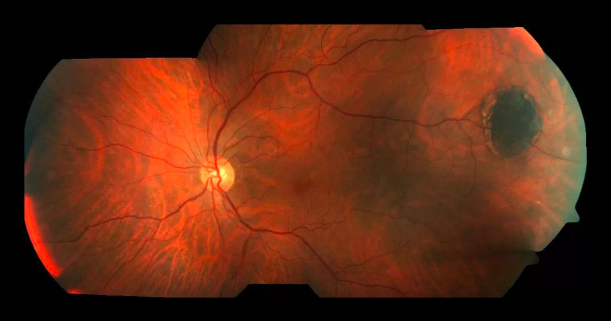
A 64-year-old Caucasian male saw his optometrist for a routine eye check. Fundus photography revealed a pigmented lesion in his left fundus.
Here you’ll find interesting cases of eye conditions along with news and developments in the ophthalmology world.
Cases are presented as an initial image with history and examination. Health practitioners are encouraged to deduce the condition, before further investigations, diagnosis and management are presented.
We hope you find it as educational, informative and exciting as we do!
Click here to view our newsletter privacy notice.
The information provided during signup is used by Eye Specialists Centre to send newsletters using the cloud-based software, Mailchimp. We do not disclose or share your personal data with other third party without your consent, or unless it is required by law. If you have any concerns about your privacy, please do not hesitate to ask.
A 64-year-old Caucasian male saw his optometrist for a routine eye check. Fundus photography revealed a pigmented lesion in his left fundus.
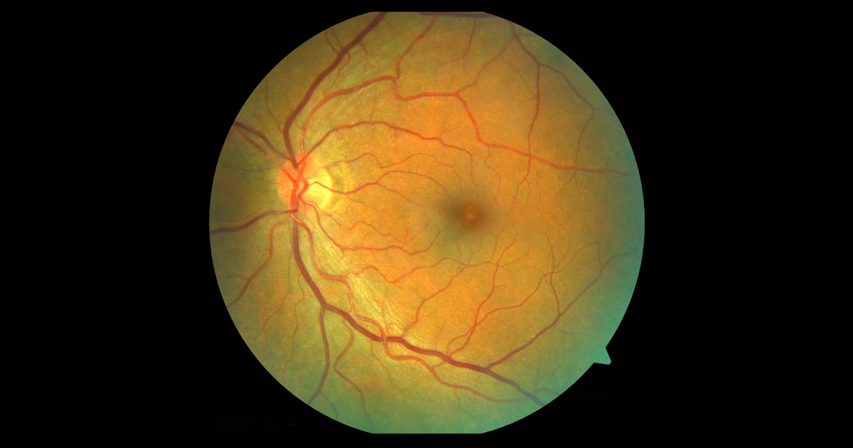
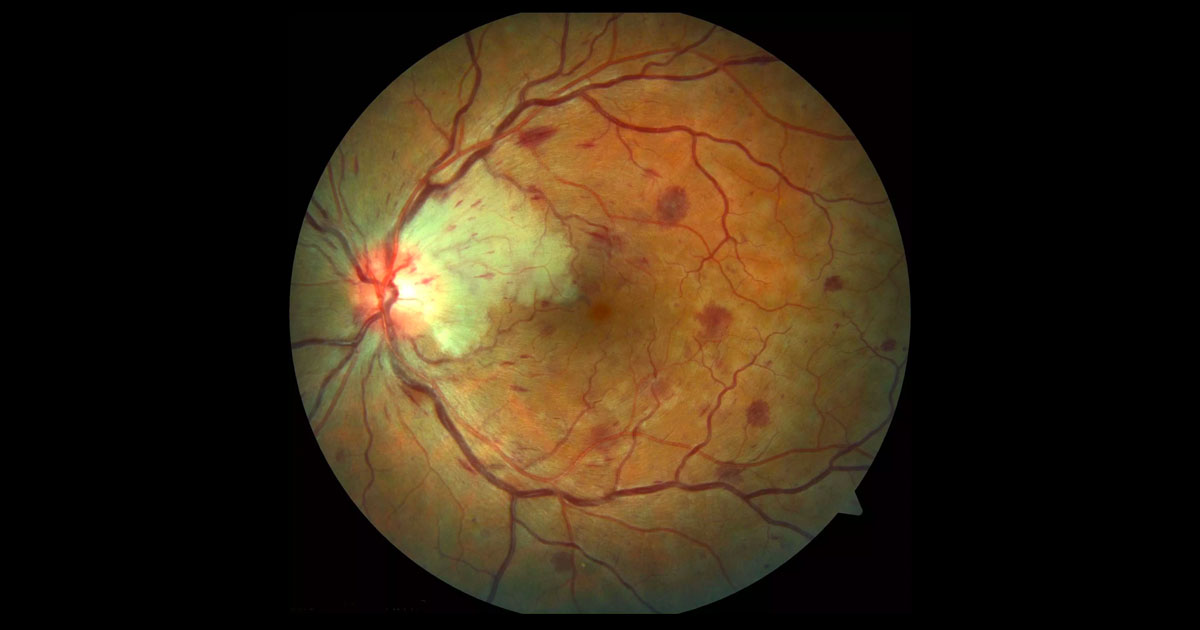
A 65-year-old male was referred with left reduced visual acuity and macular changes.
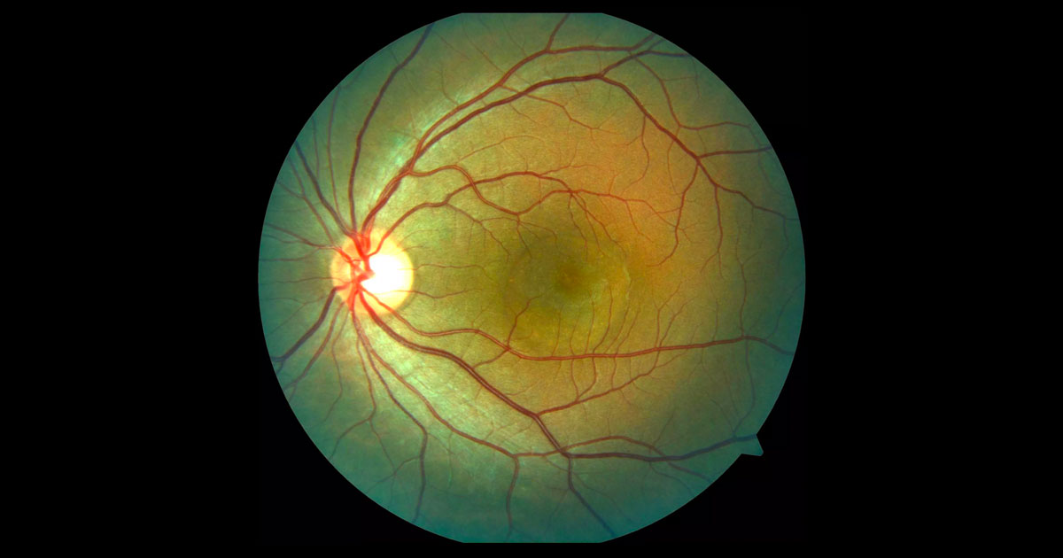
A 32-year-old man was referred with blurring of his left central vision.
A 60-year-old caucasian female was referred by her optometrist for further evaluation having presented with difficulty reading and reduced visual acuity.
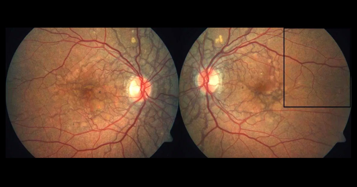
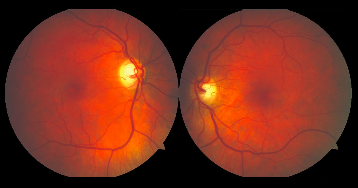
A 46-year-old Caucasian male saw his optometrist after noticing distortion in his left eye for two months. He was referred with bilateral fundus changes.
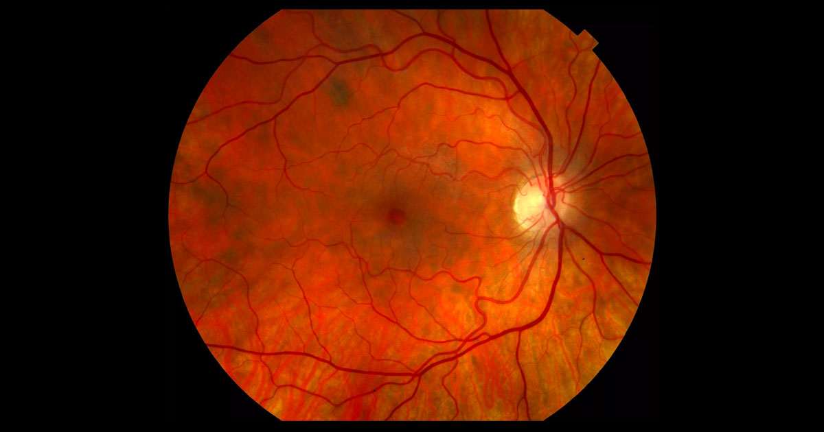
A 37-year-old lady was referred with acute painless left vision loss
A 62-year-old Russian lady was referred after her optometrist detected an irregularity at her right fovea.
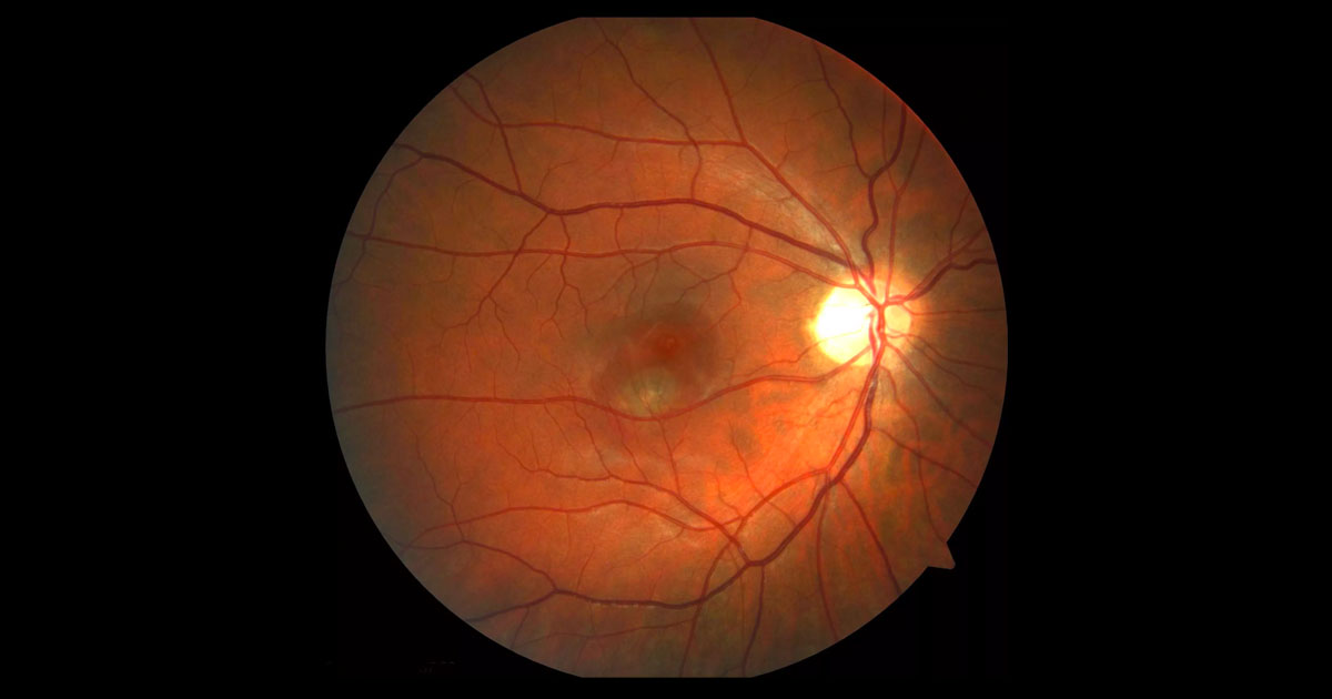
A 32-year-old nurse was referred complaining of distorted central vision in her right eye.
Have a question? Call one of our clinics today.
© 2019- Eye Specialists Centre | Privacy Policy | Disclaimer | Website design: ![]()