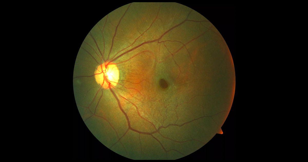
Epiretinal membrane (ERM) is a term used to describe cellular proliferation on the inner retinal surface.(1) ERMs range from benign asymptomatic thin transparent membranes through to semitransparent, thick and contractile membranes associated with debilitating metamorphopsia and central visual loss.
The majority of ERMs occur in the absence of other ocular pathology and are termed idiopathic. Secondary ERMs may occur in association with retinal vascular diseases, ocular inflammatory disease, trauma, intraocular surgery, retinal tears, detachments or laser. The most common risk factors for idiopathic ERMs include age, posterior vitreous detachment, and a history of ERM in the fellow eye.(1)
As in our case, contraction of the ERM sometimes leads to a steepening of foveal contour and a clinical appearance that resembles a macular hole, in which case the term “macular pseudohole” is used. This is a clinical diagnosis but has a characteristic OCT appearance described below. A lamellar macular hole occurs when there a defect, often splitting (schisis) of the inner retina. These terms have recently been clarified by the 2013 International Vitreomacular Traction Study Group:(2)
Macular Pseudohole
- Invaginated or heaped foveal edges
- Concomitant ERM with central opening
- Steep macular contour to the central fovea with near-normal central foveal thickness
- No loss of retinal tissue
Lamellar Macular Hole- Irregular foveal contour
- Defect in inner fovea
- Intraretinal splitting (schisis), typically between the outer plexiform and outer nuclear layers
- Maintenance of an intact photoreceptor layer
The two principle indications for surgery include reduced visual acuity and significant metamorphopsia in which case a pars plana vitrectomy and epiretinal membrane peel is performed. Removal of the internal limiting membrane is also often performed in an attempt to reduce the risk of recurrence.
(3)Surgery for lamellar macular hole alone remains controversial, with variable results. However it may be indicated when there is an associated ERM (such as in a case of macular pseudohole) and decline in vision. The majority of patients with a clinically significant ERM will respond to vitrectomy and membrane peeling. Over 80% of patients will gain two or more lines of vision and 90% report an improvement in functional vision.
(4) Typically, this improvement is gradual and observed over up to 12 months. Additionally, whilst the macular thickness often decreases after surgery the macular profile rarely returns to normal.
(5) As in our case this does not preclude satisfactory improvement of visual acuity.
(5)Photoreceptor disruption detected by optical coherence tomography has been found to be a predictor of poor visual outcome.6 Monitoring of patients with epiretinal membranes is important to detect early deterioration and intervention prior to irreversible photoreceptor damage.


