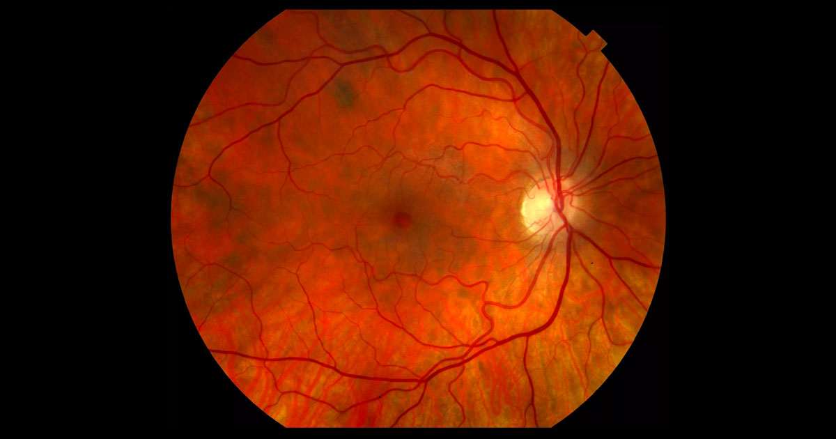
Figure 1. An ovoid cystic irregularity is present at the right fovea.
A 62-year-old Russian lady was referred after her optometrist detected an irregularity at her right fovea.
A 62-year-old emmetropic Russian lady presented to her optometrist for a routine eye check and update of her reading spectacles. An irregularity was seen at her right fovea on clinical examination. The patient was visually asymptomatic. She was referred for a further opinion.
Past ophthalmic history included left pterygium surgery. Past medical history included systemic hypertension controlled with perindopril, varicose veins and a right hip replacement. Her mother had a history of age-related macular degeneration.
Visual acuity was 6/9 (pinhole no improvement) in the right eye (OD) and 6/7.5 (pinhole 6/6-2) in the left eye (OS). Mild to moderate nuclear sclerotic and cortical cataracts were present in both eyes. An ovoid cystic irregularity was seen at the right fovea (Figure 1). There was no associated subretinal fluid, haemorrhage or lipid. Cup:disc ratio was 0.5 and the peripheral retina was normal. The vitreous was clear. A posterior vitreous detachment (with Weiss ring) was seen in the left eye but not in the right eye. The left fundus was otherwise normal.
The differential diagnosis for cystoid macular oedema includes:
- Diabetic macular oedema
- Pseudophakic cystoid macular oedema
- Retinal vein occlusion
- Age-related macular degeneration
- Vitreomacular interface pathology
- Uveitis
- Retinitis pigmentosa
Optical coherence tomography (OCT) through the right macula demonstrated the presence of vitreomacular adhesion (VMA) and an associated inner retinal cyst (Figure 2).
Figure 2. Right optical coherence tomography (horizontal raster scan) through the macula at baseline. The posterior hyaloid is detached from the retina except at the fovea, where there is an inner retinal cyst.
DIAGNOSIS
Right focal vitreomacular adhesion (VMA).
Since the patient was visually asymptomatic, a decision was made to observe closely. An Amsler grid was provided for the patient to monitor her vision. She returned 3 months later complaining of a relative central scotoma and distortion. The vision in the right eye had deteriorated to 6/12 (pinhole no improvement). Repeat OCT through the right macula showed a small full thickness macula hole (FTMH) with persistent VMA (Figure 3). As the condition was progressive and symptomatic, a decision was made to proceed with surgery.
Figure 3. Right optical coherence tomography (horizontal raster scan) through the macula three months after initial presentation. There is a small full thickness macula hole.
The patient underwent right 25-gauge vitrectomy, internal-limiting membrane peeling and sulphur hexafluoride (SF6) gas (Figure 4). She was asked to sleep on either side at night and sit up reading a book for the first 3 days.
Figure 4. Vitrectomy surgery and macular hole repair. The optic nerve is on the left and the macular hole on the right (the surgeon is sitting at the head of the patient, towards the bottom of the photograph). The internal limiting membrane has been dyed blue with Brilliant Blue G and is being peeled with forceps held in the right hand. A light pipe, held in the left hand, is illuminating the macula.
Two months following surgery, the visual acuity had improved to 6/6- OD, with an improvement in her visual symptoms. The macula hole had closed and there was restoration of the foveal architecture (Figure 5).
The patient is being followed up for post-vitrectomy cataract. She was reassured that the chances of her developing a similar problem in her left eye are low, given that she already has a complete PVD in that eye.
In youth, the posterior hyaloid (outer cortex of the vitreous) is attached to the retina. The strongest points of adhesion are at the fovea, optic disc and along retinal vessels. With age, a posterior vitreous detachment (PVD) may develop, occurring in 50% of people by 50 years and nearly all eyes by 80 years.(1) If the PVD is incomplete and points of adhesion between the vitreous and retina remain with traction, a pathological “anomalous PVD” may occur.(2)
Vitreomacular adhesion (VMA) occurs when the vitreous detaches off the retina around the macula but remains attached at the fovea. This is usually asymptomatic and non-pathologic.(3) If the vitreous causes traction at the fovea with anatomical disturbance and symptoms, the condition has progressed to vitreomacular traction (VMT). This condition may be associated with cystic changes, and patients may present with blurring of vision, a relative central scotoma and/or distortion. In addition, VMT can worsen concomitant macular disorders, in particular diabetic macular oedema and neovascular age-related macular degeneration. With further traction, VMT can in turn progress to a full thickness macular hole (FTMH).
Full thickness macular holes rarely close spontaneously and usually require treatment to improve vision. An exception is a chronic large hole when closure is unlikely to lead to visual gain due to antecedent loss of foveal photoreceptors. Currently, vitrectomy with internal limiting membrane peeling and gas endotamponade is the gold standard of treatment. Although historically a post-operative face-down posture has been advocated, there is emerging evidence that this is unnecessary for surgical success.(4) Macular hole surgery has excellent results, with successful closure over 95% of the time and improvement in vision by two lines or more in approximately 80% of patients.(5) Recently, large randomised trials have reported success of an intravitreal injection of ocriplasmin (a recombinant protease) in the treatment of VMT and small FTMHs. (6) However, this treatment is yet to become available in Australia.
TAKE HOME POINTS
- The differential diagnosis of cystoid macular oedema includes vitreomacular interface pathologies.
- Vitreomacular adhesion (VMA) occurs when the vitreous remains attached at the fovea but detached around it. There is no disturbance of the underlying macula architecture. It is asymptomatic and non-pathologic.
- Vitreomacular traction (VMT) occurs when the vitreous pulls at the fovea. It is symptomatic and pathologic. It can be associated with cystic oedema of the macula.
- Vitreomacular traction can progress to a full thickness macular hole (FTMH).
- Nearly all acute FTMHs require treatment. Vitrectomy surgery with internal limiting membrane peeling and gas is currently the gold-standard of treatment in Australia.
- There is increasing evidence that face-down positioning following vitrectomy for full thickness macula hole is not required.
- The closure rate for macula holes following vitrectomy is approximately 95%, with 80% of patients gaining two lines or more in vision.
REFERENCES
- Sebag J. Age-related changes in human vitreous structure. Graefes Arch Clin Exp Ophthalmol 1987;225:89–93.
- Sebag J. Anomalous posterior vitreous detachment: a unifying concept in vitreo-retinal disease. Graefes Arch Clin Exp Ophthalmol 2004;242:690–698.
- Stalmans P, Duker JS, Kaiser PK, Heier JS, Dugel PU, Gandorfer A, Sebag J, Haller JA. OCT-based interpretation of the vitreomacular interface and indications for pharmacologic vitreolysis. Retina 2013; 33:2003–2011.
- Iezzi R, Kapoor KG. No Face-Down Positioning and Broad Internal Limiting Membrane Peeling in the Surgical Repair of Idiopathic Macular Holes. Ophthalmology 2013;120:1998-2003.
- Lai MM, Williams GA. Anatomical And Visual Outcomes Of Idiopathic Macular Hole Surgery With Internal Limiting Membrane Removal Using Low- Concentration Indocyanine Green. Retina 2007; 27:477–482.
- Stalmans P, Benz MS, Gandorfer A, Kampik A, Girach A, Pakola S, Haller JA. Enzymatic Vitreolysis with Ocriplasmin for Vitreomacular Traction and Macular Holes. NEJM 2012. 367;7: 606-15.
Tags: cystoid macular oedema, vitreomacular traction, vitreomacular adhesion, full thickness macular hole


