Case 32
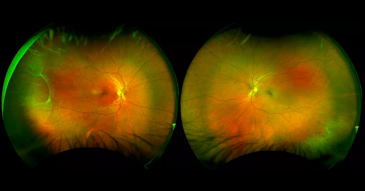
Figure 1. Colour photograph demonstrating an area of elevation in the right far temporal retina, with a fine white speckled appearance surrounding this as well as in the left inferotemporal periphery.
Author and Editor: Adrian Fung
A 43-year-old female was referred with a possible right retinal detachment.
Case history
A 43-year-old female was referred by her optometrist complaining of a one-month history of flashes and floaters in her right eye. She also complained of headaches starting that day. The optometrist was concerned she had rhegmatogenous retinal detachment. There was no previous ophthalmic history nor any relevant medical history.
Visual acuity was 6/9.5+2 ph 6/4.8-1 in the right eye (OD) and 6/6 in the left eye (OS). Intraocular pressures were 21mmHg (OD) and 19mmHg (OS). Anterior segment examination was normal and there was no vitreous pigmentation. On fundus examination there was an area of elevation between the 8 and 10 o’clock meridia in the righ eye with a fine white speckled appearance (Figure 1). A similar fine white speckled appearance was detected in the left inferotemporal periphery, but without any elevation. Both maculae and optic nerves were normal. The posterior hyaloids were still attached.
