Case 48
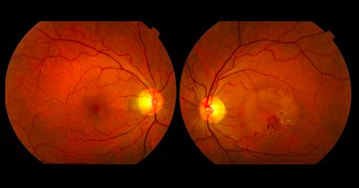
Figure 1. Colour fundus photographs shows a circular area of macular elevation associated with haemorrhage and hard exudate in the left eye.
Author: Raj Chalasani Editor: Adrian Fung
A 54-year-old male was referred with reduced vision in his left eye.
Case history
A 54-year-old male was referred by his optometrist with a 2-month history of blurring of vision in his left eye, most noticeable over the previous 2 weeks.
Best corrected visual acuities were 6/5 in the right eye (OD) and 6/12 in the left eye (OS). Intraocular pressures were 15mmHg in both eyes. Examination of the anterior segment was unremarkable. Examination of the posterior segments revealed a circular area of macular elevation associated with haemorrhage and hard exudate in the left eye, and pigmentary changes in the right eye (Figure 1). No drusen were visible.

