Case 5
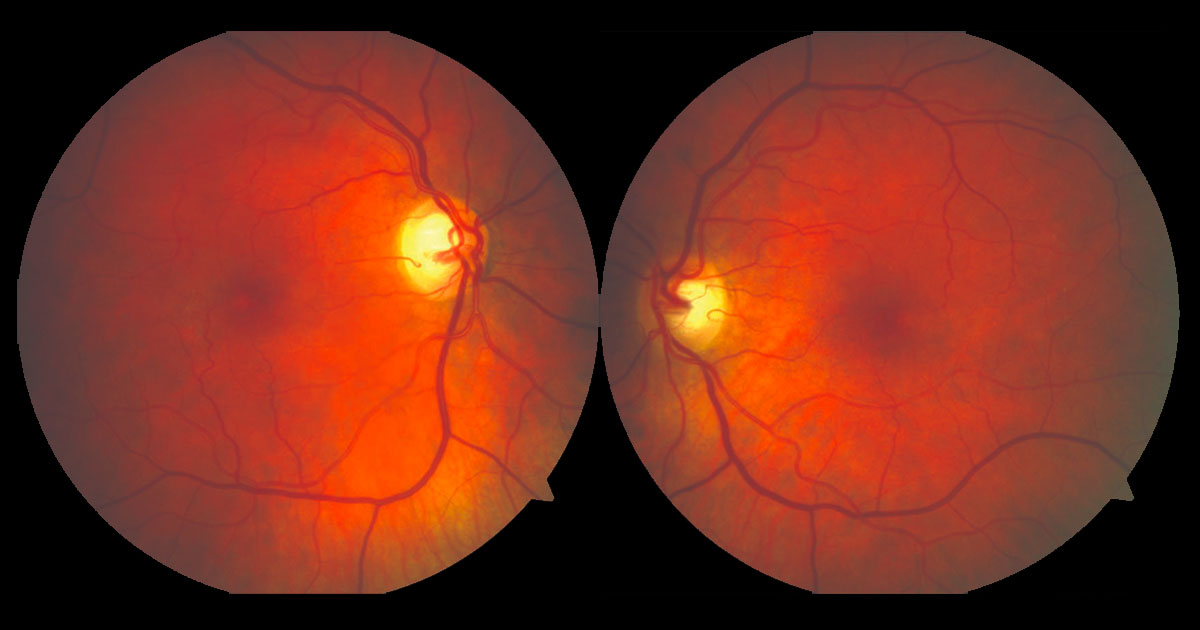
Figure 1a. Colour fundus photographs reveal subtle abnormalities temporal to both foveae.
Author: James Leong Editor: Adrian Fung
A 60-year-old caucasian female was referred by her optometrist for further evaluation having presented with difficulty reading and reduced visual acuity.
Case history
A 60-year-old caucasian female was referred with complaints of a gradual reduction in her visual acuity and difficulty reading over the last 12 months. She had no significant past ocular history although there was a family history of glaucoma. Past medical history included diet controlled diabetes mellitus and hypertension.
On examination her visual acuities were 6/18 pinhole 6/12 in the right eye (OD) and 6/7.5 (pinhole no improvement) in the left (OS). Intraocular pressures were 16mmHg OD, 14mmHg OS. Anterior segment examination was normal. Examination of both maculae demonstrated loss of the foveolar reflex and reduced retinal transparency, yellow crystalline spots and mild intraretinal pigment hyperplasia. Mildly telangiectatic capillaries with right angle venules were evident temporal to the foveae (Figs 1a and b).

Figure 1b. Magnified image of the right macula demonstrates the following changes temporal to the fovea: reduced retinal transparency, yellow crystals, intraretinal pigmentation and telangiectatic vessels with right angle venules.


