Case 8
Author and Editor: Adrian Fung
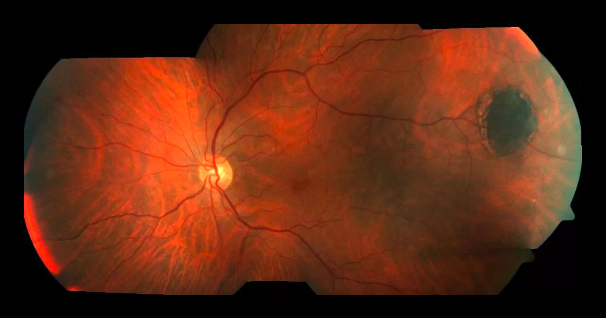
Figure 1a. Colour montage fundus photograph of the left eye reveals a flat pigmented lesion at the temporal equator.
A 64-year-old Caucasian male saw his optometrist for a routine eye check. Fundus photography revealed a pigmented lesion in his left fundus.
Case history
A 64-year-old Caucasian male saw his optometrist saw his optometrist for a routine eye check. Wide-field fundus photography revealed a left pigmented fundus lesion.
The patient had previously undergone uneventful LASIK surgery for myopia. He was otherwise well and there was no history of cutaneous melanoma or systemic carcinoma. He last smoked 18 years prior. There was no family history of ocular tumours.
Visual acuity was 6/6-2 in the right eye (OD) and 6/9.5-2 (pinhole 6/4.8-1) in the left eye (OS). On examination there was no ocular melanocytosis, iris heterochromia or cervical lymphadenopathy. Anterior segments were normal and both irides were brown. On fundus examination there was a flat, round, pigmented fundus lesion in the left temporal equator (Figures 1a and b). This measured 4.0X3.0mm and had a nasal halo of depigmentation. There was no associated orange pigment, subretinal fluid or drusen.

Figure 1b. Magnified view of the left temporal pigmented lesion.


