Case 15
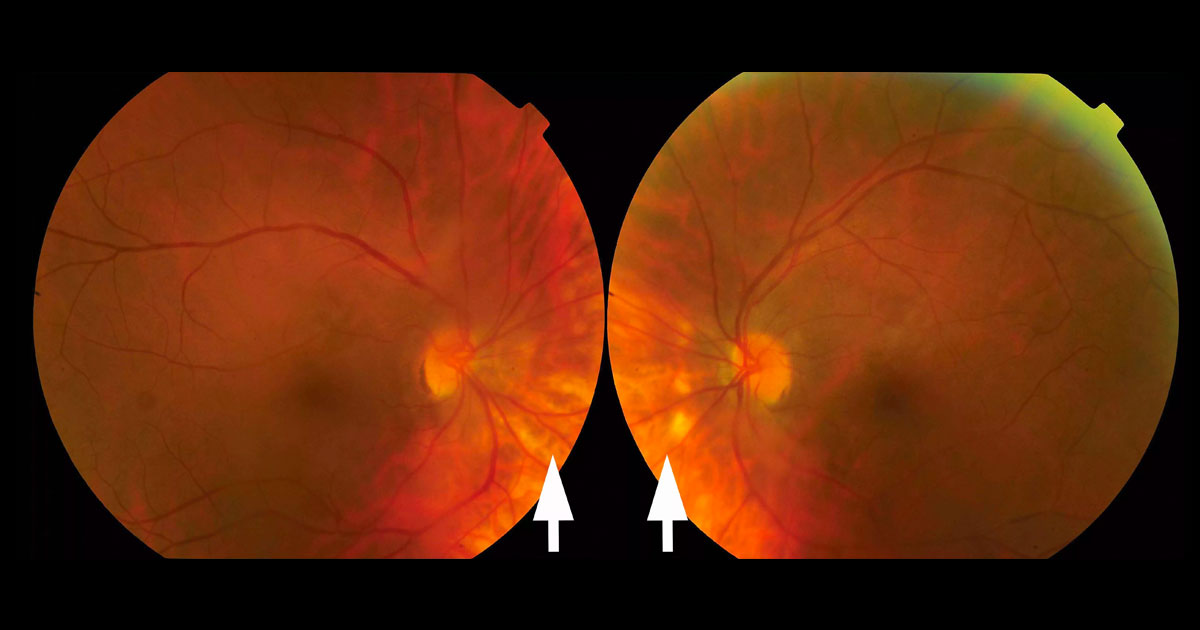
Figure 1. Colour fundus photographs reveal multiple, deep, yellow-white lesions nasal to the optic discs.
Author: Raj Chalasani Editor: Adrian Fung
A 54-year-old Caucasian female saw her optometrist complaining of bilateral flashes and floaters.
Case history
A 54-year-old Caucasian female saw her optometrist complaining of bilateral photopsiae and floaters. The patient was otherwise well and there was no previous significant ocular history.
Visual acuity was 6/7.5 in the right eye (OD) and 6/6-2 in the left eye (OS). Intraocular pressures were normal. Anterior chambers were quiet, but non-pigmented vitreous cells were present bilaterally. On fundus examination multiple, deep, yellow-white lesions were visible in the posterior pole, most prominent nasal to the optic discs.

