Case 41
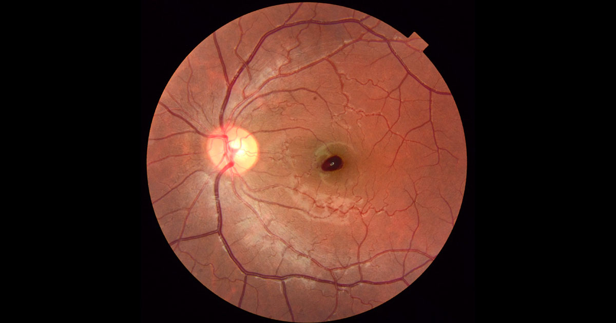
Figure 1. Colour fundus photograph of the left eye shows a well-circumscribed dark patch at the fovea.
Author: Simon Nothling Editor: Adrian Fung
A 21-year-old female was referred with a dark patch at her left macula.
Case history
A 21-year-old professional female rugby player presented with a 1-day history of painless vision loss and central scotoma in her left eye. She had no other neurological symptoms such as headache or limb paraesthesiae. The day before she had been performing some intense scrummaging at training, as well as some heavy weight-lifting, which was slightly more than normal. She denied any coughing or other straining recently. She had no ophthalmic history and was well medically. She denied being pregnant.
On examination, her visual acuities were 6/5-1 right eye (OD) and 6/24-1 left eye (OS) unaided, with no improvement with pinhole. Intraocular pressures were 15mmHg (OD) and 16mmHg (OS). Anterior segment examination was normal in both eyes. Blood pressure was 120/75. Colour fundus photographs reveal a left well-circumscribed, dome-shaped dark patch at the fovea (Figure 1). The right fundus was normal.


