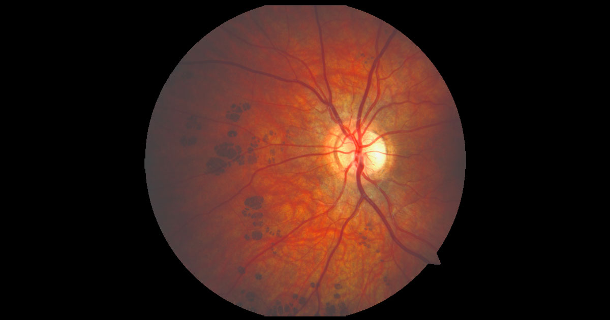
Figure 1. Colour fundus photograph of the left eye demonstrates well defined flat pigmented lesions nasal to the optic nerve.
A 14-year-old boy was referred by his optometrist with unusual areas of fundus pigmentation in his left eye.
A 14-year-old boy was referred by his optometrist, who had noted areas of abnormal fundus pigmentation in his left eye. His ophthalmic history was unremarkable and this had been his first formal eye examination. There was a past medical history of mild eczema.
Visual acuity was 6/6 in the right eye (OD) and 6/6- in the left eye (OS). The anterior segment examination was unremarkable. There was no cataract. Intraocular pressures were OD 14mmHg and OS 15 mmHg. Fundus examination of the left eye demonstrated groups of well-defined areas of pigmentation throughout the retina in the left eye (Figure 1). These were not elevated and blood vessels could be seen running smoothly over the top of some of the lesions.
The causes for multiple areas of fundus hyperpigmentation include:
- Congenital hypertrophy of the retinal pigment epithelium (CHRPE)
- Acquired hypertrophy of the RPE
- Iatrogenic- previous retinal argon laser treatment/ panretinal photocoagulation
- Previous trauma
- Previous infection (e.g. syphilis, rubella)
- Multifocal choroidal naevi
- Retinitis pigmentosa
Fundus autofluorescence demonstrated the lesions to be homogenously hypoautofluorescent (Figure 2).
Figure 2. Left fundus autofluorescence demonstrates the lesions to be homogenously hypoautofluorescent.
DIAGNOSIS
Grouped congenital hypertrophy of the retinal pigment epithelium (CHRPE).
Congenital hypertrophy of the retinal pigment epithelium (CHRPE) are benign congenital lesions. They are usually asymptomatic and an incidental finding on dilated fundus examination.
CHRPE present as flat, pigmented, well-defined fundus lesions at the level of the retinal pigment epithelium (RPE).(1) The most common clinical phenotype is of solitary CHRPE. They are well demarcated, often having a depigmented halo which outlines the lesion. With time lesions may will slowly grow in diameter.(2) Many CHRPE also have areas of depigmentation within the lesion known as lacunae. The number and size of lacunae typically increases over time, and may fill the entire lesion. CHRPE lesions have rarely been reported to undergo nodular transformation due to the formation of a primary malignant adenocarcinoma of the RPE.(3) The lesions are typically hypoautofluorescent, with outer retinal loss on optical coherence tomography.(4) This can be useful if the diagnosis is uncertain.
The other less common fundus phenotype is of grouped CHRPE lesions, as seen in this case. These lesions are multiple, small, brown-black in colour, and often resemble footprints, explaining the colloquial name “bear tracks”. Usually such bear tracks are larger in the periphery and decrease in size towards the optic disc (Figure 3).
Grouped CHRPE need to be distinguished from the pigmented fundus lesions seen in association with familial adenomatous polyposis (FAP), a familial cancer syndrome associated with colorectal cancers. Pigmented fundus lesions are the most common extra-colonic manifestation of FAP, occurring in approximately 60% of patients.(5) The lesions associated with FAP are different from typical CHRPE in that they are usually bilateral, multiple, oval or fish shaped in appearance with a depigmented fishtail (Figure 4).
Figure 3. Left colour and autofluorescence images of the posterior pole demonstrating the lesions to be less prominent and smaller.
Figure 4. A fundus lesion typical of those associated with familial adenomatous polyposis. There is a characteristic depigmented “fishtail”.
TAKE HOME POINTS
- Grouped CHRPE are congenital benign lesions of the retinal pigment epithelium, sometimes described as “bear tracks”.
- They appear as flat, well-defined brown-black lesions fundus lesions.
- They are hypoautofluorescent and exhibit overlying outer retinal thinning.
- They should be distinguished from the pigmented fundus lesions associated with familial adenomatous polyposis. These are multiple, bilateral, oval in shape and have a depigmented “fishtail”.
REFERENCES
- Gass JD. Focal congenital anomalies of the retinal pigment epithelium. Eye 1989;3:1–18.
- Arepalli S, Kaliki S, Shields JA, Shields CL. Growth of congenital hypertrophy of the retinal pigment epithelium over 22 years. J Pediatr Ophthalmol Strabismus. 2012 Dec 18;49 Online:e73-5
- Shields JA, Eagle RC Jr, Shields CL et al Malignant transformation of congenitalhypertrophy of the retinal pigment epithelium Ophthalmology. 2009 Nov 116(11):2213-6.
- Fung AT, Pellegrini M, Shields CL. Congenital Hypertrophy of the Retinal Pigment Epithelium. Enhanced-depth imaging optical coherence tomography in 18 cases. Ophthalmology 2014; 121:251-256
- Groen EJ Roos A, Munthinge FLet al. Extra-Intestinal Manifestations of Familial Adenomatous Polyposis. Ann Surg Oncol. 2008 Sep; 15(9): 2439–2450.
Tags: fundus pigmentation, chrpe, familial adenomatous polyposis, vision loss


